A Real-time Image Stitching Framework for Fetoscopic Field-of-view Expansion
Rowan Honeywell, Radian Gondokaryono, Rory Windrim, Lueder A. Kahrs,
Medical Computer Vision and Robotics Lab, University of Toronto
Abstract
Objectives: Twin-to-twin transfusion syndrome is a serious prenatal condition whose treatment employs the use of a fetoscope and optical laser for targeted vessel ablation. During surgery, challenges such as a limited field-of-view and inconsistent lighting can be addressed by field-of-view expansion.
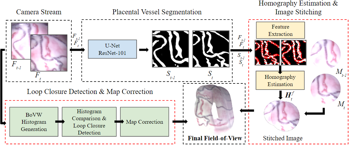
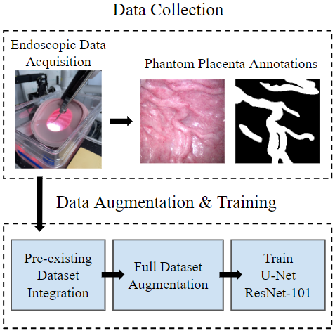
Methods: As foundation, we draw upon a state-of-the-art method that utilizes a U-Net segmentation model to mitigate visible in utero inconsistencies. We adapt this method for real-time performance by implementing a feature-based stitching algorithm. The proposed framework includes a loop closure and map correction procedure to help reduce the accumulated drifting error.
Results: During evaluation on simulated fetoscopic movements, the chosen feature extractor, SIFT, permits an average homography estimation time of 0.07s and a failure rate of 4%. Homography parameters such as scaling, translation, and rotation are isolated to identify obstacles caused by certain camera movements. The framework is evaluated using two phantom placentas in different environmental conditions and submerged underwater. Qualitative and quantitative results are presented as well as an analysis of the map correction procedure. These evaluations help recognize limitations of the framework that can be investigated and addressed in future research.
Conclusion: Deep segmentation of placental vessels has been shown to increase image stitching accuracy which, in turn, can improve the quality of TTTS treatment. Our proposed framework builds upon this concept by introducing a feature-based (SIFT) homography estimation to enable real-time image stitching at a rate of at least 10 frames per second. To help balance the speed-accuracy trade-off, the proposed framework also incorporates a simple loop closure-based map correction procedure. Quantitative testing justified the use of SIFT over other popular feature extractors due to its reliability and compatibility with real-time use. Qualitative testing showed that the framework performs consistently across various simulated and in vivo environments.


BibTeX Citation
  @InProceedings{honeywell2024fetoscopicfov,
	author={Honeywell, Rowan
	and Gondokaryono, Radian
	and Windrim, Rory
	and Kahrs, Lueder A.},
	title={A Real-Time Image Stitching Framework for Fetoscopic Field-of-View Expansion},
	booktitle={21st International Symposium on Biomedical Imaging},
	year={2024},
	publisher={IEEE},
	pages={tbd},
	doi={tbd}}
